Radius Bone Labelled Diagram : The Hard Working Foot, Part One - Chicago Dance Supply : The following pages may be of.. Proximal radius (head, neck and tuberosity). The bones shown in the chest and hip region in the labeled human skeleton diagram are the ribs humerus is located in the upper arm. The radius bone is shorter. Learn radius and ulna anatomy with these fun quizzes and diagrams. 12 photos of the labelled diagram of radius bone.
The radius is considered the most commonly fractured bone in the human body, with distal radius fractures being the most common form of radial. The lateral side projects distally as the styloid process. Skull, clavicle, mandible, scapula, thorax, sternum, humerus, ulna, radius, carpus, phalanges (fingers), metacarpus, spine, pelvis, sacrum, femur, tibia, fibula, tarsus. A basic human skeleton is studied in schools with a simple diagram. For more information about the adjacent this website includes many pages about bones and the skeletal system.
Gross anatomy of commonly fractured bones from bonefracturerepair.webnode.com The radius and ulna together constitute the forearm. Each bone is a complex living organ that is made up of many cells, protein fibers, and minerals. It is one of the two bones of the forearm, the other being the ulna. For more information about the adjacent this website includes many pages about bones and the skeletal system. The biceps originate near the shoulder joint and insert into the radial tuberosity on the upper part of the radius, near the elbow joint. Study guide for students and teachers. The radius bone is this bone here and it lies laterally in the anatomical position. Labeled ulna and radius » diagram text anterior labeled ulna and radius anatomy of the ulna and radius bones print options de labeled ulna.
The radius and ulna together constitute the forearm.
All land vertebrates have this bone. Radius definition location functions anatomy diagram. Proximal radius (head, neck and tuberosity). In its distal part, the radial shaft expands to form a rectangular end. A basic human skeleton is studied in schools with a simple diagram. The lateral side projects distally as the styloid process. Knowledge of the location the location of the radius is indicated in the diagram. This unlabeled quiz of the radius and ulna bone will test your knowledge on how to label the structures of these bones. Skull, clavicle, mandible, scapula, thorax, sternum, humerus, ulna, radius, carpus, phalanges (fingers), metacarpus, spine, pelvis, sacrum, femur, tibia, fibula, tarsus. Projection of bone on the lateral surface of the distal radius bone. The following pages may be of. 6 3 bone structure u2013 anatomy and physiology. The photograph may be purchased as wall art, home decor, apparel, phone cases, greeting cards, and more.
Its upper concave surface articulates with the. Skeletal system quizzes learn bone anatomy fast. Exam 2 bones of the lower limb. It extends from the lateral side of the elbow to the thumb side of the wrist and runs parallel to the ulna. The bones mentioned in each human skeleton chart are:
MD 3 Anatomy - Anatomy 2017 with Sloz at John A. Burns ... from classconnection.s3.amazonaws.com The ulna articulates with the trochlea and the radius articulates with the capitulum. Radius bone diagram labeled ulna bone anatomy bones of the hand labeled new bones of the upper limb in. Skeletal system quizzes learn bone anatomy fast. The radius is the home for a few muscles' insertion points. The biceps originate near the shoulder joint and insert into the radial tuberosity on the upper part of the radius, near the elbow joint. Radius definition location functions anatomy diagram. Each bone is a complex living organ that is made up of many cells, protein fibers, and minerals. The radial head of the flexor digitorum superficialis takes origin from the anterior oblique line and upper part of the anterior border.
The biceps originate near the shoulder joint and insert into the radial tuberosity on the upper part of the radius, near the elbow joint.
In the diagram of the tibia and fibula, this structure articulates with the talus and forms a protrusion on the medial surface of the ankle. Radius along with ulna connects elbow to forearm. Learn vocabulary, terms and more with flashcards, games and other study tools. Radius ulna bones ulnar notch anterior anatomy markings head forearm wrist hand limb upper pronation pivot supination during around. Each bone is a complex living organ that is made up of many cells, protein fibers, and minerals. The radius and ulna are the two bones of the forearm. Short video describing the skeletal structures of the radiusstructures identified:headneckradial tuberositystyloid process of the radiusulnar notch. The lateral side projects distally as the styloid process. There are different features on each bone that also can help distinguish between the. Study guide for students and teachers. 4 lower end presents a tubercle on the posterior surface called as dorsal tubercle of lister. Thats the way i remembered which bone is located in anatomy class. This ulnar view labelled illustration is from 'asklepios atlas of the human anatomy'.
In the diagram of the ulna and radius, where is the radial tuberosity? For more information about the adjacent this website includes many pages about bones and the skeletal system. The radius bone is a long horizontal bone present in the forearm and is also called the radial bone. Definition, location, functions, anatomy, diagram. The two bones play only secondary roles at their opposing joints.
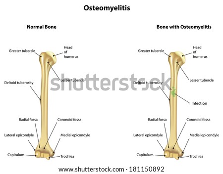
Osteomyelitis Labeled Diagram Humerus Bone Stock ... from image.shutterstock.com Even my bio teacher get confused. Short video describing the skeletal structures of the radiusstructures identified:headneckradial tuberositystyloid process of the radiusulnar notch. Labeled ulna and radius » diagram text anterior labeled ulna and radius anatomy of the ulna and radius bones print options de labeled ulna. Close to neck it presents a radial tuberosity. Learn radius and ulna anatomy with these fun quizzes and diagrams. All land vertebrates have this bone. It extends from the lateral side of the elbow to the thumb side of the wrist and runs parallel to the ulna. The two bones play only secondary roles at their opposing joints.
It extends from the lateral side of the elbow to the thumb side of the wrist and runs parallel to the ulna.
Definition, location, functions, anatomy, diagram. Thats the way i remembered which bone is located in anatomy class. Its upper concave surface articulates with the. Close to neck it presents a radial tuberosity. Radius bone is a photograph by asklepios medical atlas which was uploaded on august 3rd, 2016. Study guide for students and teachers. Proximal radius (head, neck and tuberosity). Radius definition location functions anatomy diagram. 769 x 1000 jpeg 102kb. 800 x 701 jpeg 80kb. Exam 2 bones of the lower limb. Labeled ulna and radius » diagram text anterior labeled ulna and radius anatomy of the ulna and radius bones print options de labeled ulna. The following pages may be of.
For more information about the adjacent this website includes many pages about bones and the skeletal system labelled radius bone. It extends obliquely downward into a strong, conical projection.


0 Komentar